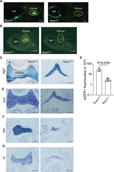
Impaired intrathymic lymphopoiesis in foxn1-deficient fish. (A) Whole mount depiction of zebrafish at 6 dpf, transgenic for an ikzf1:eGFP pan-lymphocyte reporter transgene. The foxn1 genotypes are indicated, as are anatomical landmarks; note that subpopulations of neurons also express ikzf1. Scale = 0.1 mm. (B) Macroscopic appearance of adult fish (6 weeks of age) transgenic for the ikzf1:eGFP reporter transgene. The foxn1 genotypes are indicated. Scale = 1 mm. (C) RNA in situ hybridization of thymic tissue sections using a rag1-specific probe. Note that the medulla (rag1-negative region) is barely visible in the mutants. (D) RNA in situ hybridization of thymic tissue sections using a tcrb-specific probe. (E) Adult mutant fish (6 weeks of age) possess fewer eGFP-positive thymocytes as determined by flow cytometry on cells isolated from microdissected thymic lobes (mean ± s.e.m. are shown; t-test, two-tailed). Each data point represents an individual fish. (F) RNA in situ hybridization of thymic tissue sections using a dll4-specific probe. (G) RNA in situ hybridization of thymic tissue sections using an il7-specific probe. For C, D, F, G, scale, 0.1 mm.
|
