
Fig. 6
- ID
- ZDB-FIG-240219-14
- Publication
- Murali et al., 2024 - Genetic variant classification by predicted protein structure: A case study on IRF6
- Other Figures
- All Figure Page
- Back to All Figure Page
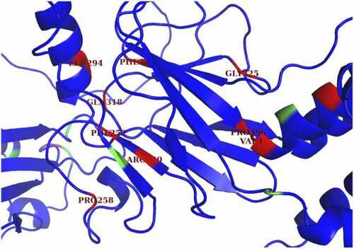
Potential visible binding pocket in protein-binding domain. This image shows a zoomed-in version of the AlphaFold2 structure of full IRF6. Some of the residues in or near the beta sheet, which is mainly part of the protein-binding domain, were categorized as ruptured. Although some of these residues are distant from each other in the amino acid sequence, there are some pairs that are close together in the 3D folded structure, such as ARG-250 and PHE-375. The clustering of red mutations in the protein-binding domain highlights the significance of this domain for IRF6 protein function and provide insight on which proteins may be more likely to be deleterious. |