
Fig. 1
- ID
- ZDB-FIG-240216-10
- Publication
- Canato et al., 2023 - Anti-HER2 Super Stealth Immunoliposomes for Targeted-Chemotherapy
- Other Figures
- All Figure Page
- Back to All Figure Page
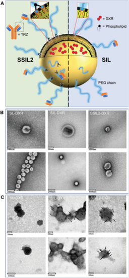
Schematic representation and TEM images of liposomes. A) Drawing of SSIL2 and SIL highlighting the different anchoring of PEG chains on the phospholipid bilayer. B) TEM characterization of SL-DXR, SIL-DXR, and SSIL2-DXR. C) Immunolabeling of targeted liposomes (SIL and SSIL2) compared to non-targeted liposomes (SL) as the negative control. The black dots indicated by the arrows correspond to the gold nanoparticles (5 nm) conjugated to a secondary anti-mouse antibody binding the primary anti-Trastuzumab antibody, thus allowing indirect visualization of the Fab’ fragments on the surface of liposomes. The gold nanoparticles were not detected in non-targeted liposomes. |