|
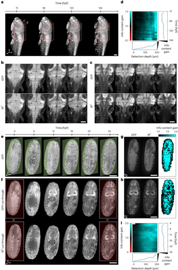
Infrared-mediated image restoration provides high-contrast deep-tissue time-lapse imaging of living biological systems. a, 3D reconstruction of live Tg(h2b:gfp) zebrafish larva images. Orange boxes represent the regions shown in b,c. Scale bar, 100 µm. b,c, Individual z planes at detection depth of 100 µm and 250 µm, respectively for the fish larva shown in a. Endogenous GFP (top), IR2 reconstructed images (bottom). Scale bar, 100 µm. d, Kymograph representing the information content gain relative to the GFP images, for all the images in the time-lapse dataset and as a function of detection depth. Line plots to the right and the bottom represent the depth- and time-average information content gain. e, 3D reconstruction of live (Tg(His2AV-GFP)) fly larva images. Green opaque planes represent the sample sections shown in f. Scale bar, 100 µm. f, Individual z plane for the fly images shown in e. Endogenous GFP (top), IR2 reconstructed images (bottom). Red-highlighted time points are shown in g,h. Scale bar, 100 µm. g,h, Spatial mapping of the information content gain for the two individual z planes shown. Scale bar, 100 µm. i, Kymograph of the information content gain as a function of time and detection depth. Line plots represent the depth- and time-average information content gain.
|