|
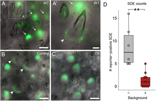
Bmp6 OE caused a reduction of TCF/Lef reporter activity in successional dental epithelia. (A,A′) Images of WT pharyngeal tooth fields with a TCF/Lef reporter transgene. Arrows mark tooth germs, arrowhead marks successional dental epithelium. A′ shows an enlarged region of A as marked by the white dotted box. (B,C) Control (B) and OE (C) example images of pharyngeal teeth with the same reporter construct. Note the GFP-positive successional dental epithelia in the control individual (arrowheads). (D) A box and whisker plot showing the number of TCF/Lef-positive successional dental epithelia (SDE). Boxes represent the 25th-75th percentiles, the median is shown as a black bar, and whiskers represent those data within 1.5× the interquartile range above and below the 75th and 25th percentiles, respectively. Wilcoxon Rank-Sum test, **P<0.01 (P=0.0021; n=6 control, 9 OE fish). Scale bars: 100 μm (A-C); 25 μm (A′).
|