IMAGE

Fig. 8.

ID
ZDB-IMAGE-231220-49
Source
Figures for Square et al., 2023
Image
Figure Caption

Fig. 8.

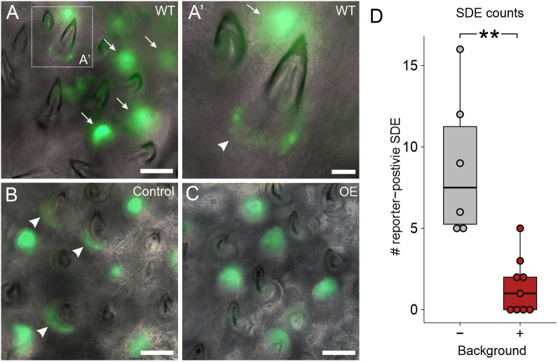
Bmp6 OE caused a reduction of TCF/Lef reporter activity in successional dental epithelia. (A,A′) Images of WT pharyngeal tooth fields with a TCF/Lef reporter transgene. Arrows mark tooth germs, arrowhead marks successional dental epithelium. A′ shows an enlarged region of A as marked by the white dotted box. (B,C) Control (B) and OE (C) example images of pharyngeal teeth with the same reporter construct. Note the GFP-positive successional dental epithelia in the control individual (arrowheads). (D) A box and whisker plot showing the number of TCF/Lef-positive successional dental epithelia (SDE). Boxes represent the 25th-75th percentiles, the median is shown as a black bar, and whiskers represent those data within 1.5× the interquartile range above and below the 75th and 25th percentiles, respectively. Wilcoxon Rank-Sum test, **P<0.01 (P=0.0021; n=6 control, 9 OE fish). Scale bars: 100 μm (A-C); 25 μm (A′).

Acknowledgments
This image is the copyrighted work of the attributed author or publisher, and ZFIN has permission only to display this image to its users. Additional permissions should be obtained from the applicable author or publisher of the image. Full text @ Development