|
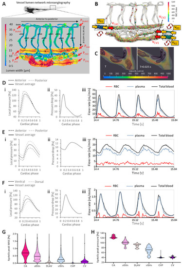
Development of the cell-and-plasma 3-D computational fluid dynamics (CFD) model (A) Morphology of zebrafish trunk network in the caudal vein plexus region obtained using microangiography and confocal microscopy. Shown are the different vessel types of varying diameter (CA: caudal artery, CV: caudal vein, aISV and vISV: arterial and venous intersegmental vessels, DLAV: dorsal longitudinal anastomotic vessel, CVP: caudal vein plexus). Red arrows indicate the 7 locations where pulsatile pressure input boundary conditions are specified. (B) Red blood cell (RBC) hematocrit in the simulation domain controlled and maintained by recycling cells in the 3 dashed-box domains (RBC indices < 344), see S1 Movie. Yellow arrows indicate reservoir velocity periodic boundary inputs copied from the adjacent region (red arrow) in the trunk network. (C) Collision of RBCs with vessel walls and with one another as part of the explicit consideration of blood dynamics in the CFD model (S2 Movie). (D and E) Pulsatile pressure boundary conditions defined at the anterior and posterior ends of the CA (Di) and CV (Ei) and the resulting pulsatile pressure drop across the CA (Dii) and CV (Eii) that drives the pulsatile flow of RBC and plasma blood phases in the CA (Diii) and CV (Eii). (F) Pulsatile pressures arising at the dorsal and ventral ends of aISV3 (i) due to oscillating pressure inputs at anterior and posterior ends of the CA and CV, and the resulting pulsatile pressure drop across aISV3 (ii) that drives the pulsatile flow of RBC and plasma blood phases in aISV3 (iii). (G and H) Hierarchical stratification of WSS (G) and blood pressure (H) levels in the network predicted by CFD; see S1 Movie for spatial maps of oscillating WSS and pressure.
|