FIGURE

Figure 7

ID
ZDB-FIG-231002-442
Publication
Karolczak et al., 2023 - Loss of Mtm1 causes cholestatic liver disease in a model of X-linked myotubular myopathy
Other Figures
All Figure Page
Back to All Figure Page
Figure 7

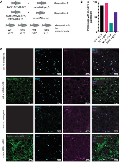
Liver-specific Mtm1 expression rescues the cholestatic phenotype of mtm zebrafish.

The fabp:mtm1-GFP transgene was introduced into the mtm-mutant zebrafish line. The resulting fish were analyzed for morphological and functional changes associated with cholestasis. (A) Crossing scheme for introducing the fabp:mtm1-gfp transgene into the mtm zebrafish line. Transgenic fish were outcrossed twice to mtm1+/Δ8 fish, resulting in clutches of larvae for experiments that contained WT and mtm fish with and without the transgene. (B) A BODIPY assay was used to measure bile flux (WT – GFP = 88%, WT + GFP = 96%, mtm – GFP = 30%, mtm + GFP = 65%). In the mtm transgene–positive group, there were more mtm mutants with normal bile flux when compared with the mtm transgene–negative group, as measured by positive gall bladder fluorescence (P = 0.0532, 1-sided Fisher’s exact test). (C) Visualization of liver-specific Mtm1 expression from the fabp:mtm1-GFP transgene. In WT fish, Mtm1 localized to the plasma membrane and to subapical structures that were Mdr1+ and Rab11+ by immunostaining (top two rows). In mtm zebrafish, hepatocyte-expressed Mtm1 restored bile canalicular architecture and bile transporter expression to the bile canaliculi. Coimmunostaining of whole-mount embryos revealed the reexpression of Mdr1 puncta in 7 dpf mtm larvae. Scale bars: 10 μm.

Expression Data

Expression Detail
Antibody Labeling
Phenotype Data

Phenotype Detail
Acknowledgments
This image is the copyrighted work of the attributed author or publisher, and ZFIN has permission only to display this image to its users. Additional permissions should be obtained from the applicable author or publisher of the image. Full text @ Journal of Clin. Invest.