|
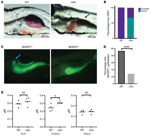
mtm zebrafish show evidence of hepatic steatosis and cholestasis. (A) Oil Red O staining shows hepatic steatosis in 5 dpf zebrafish larvae. Dashed lines outline the liver, where there is evidence of lipid accumulation in mtm zebrafish. Black arrow denotes the swim bladder, which is not properly inflated in mtm mutants. Scale bars: 100 μm. (B) Quantification of Oil Red O in liver (n = 10 fish for each condition). No WT larvae had severe steatosis compared with 66% of mtm larvae. (C) BODIPY feeding assay showed impaired bile flux in mtm larvae. WT and mtm zebrafish were fed AP-100 fish food mixed with BODIPY C12. Representative images are shown of fish with positive and negative gallbladder fluorescence. Blue arrow denotes the gallbladder. Original magnification, ×25. (D) Quantification of gallbladder fluorescence combining 2 independent biological replicates (n = 30 fish per replicate). Ninety-three percent of WT larvae exhibited gallbladder fluorescence after BODIPY exposure, whereas only 28% of mtm larvae showed gallbladder fluorescence (****P < 0.0001, by Fisher’s exact test). (E) Comparisons of individual bile acids at 5 dpf. TCA, TCDCA, and TDCA are 3 conjugated, hydrophobic bile acids that can cause toxicity when their levels are elevated. TCA and TDCA levels appeared unchanged, whereas TCDCA levels were elevated in mtm larvae (*P = 0.039, by unpaired, 2-tailed t test).
|