FIGURE

Figure 2

ID
ZDB-FIG-231002-437
Publication
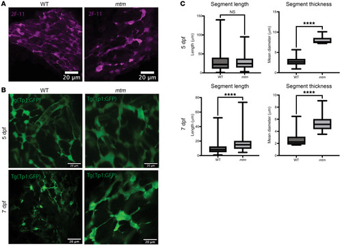
Karolczak et al., 2023 - Loss of Mtm1 causes cholestatic liver disease in a model of X-linked myotubular myopathy
Other Figures
All Figure Page
Back to All Figure Page
Figure 2

mtm zebrafish have dilated bile ducts with reduced branching.

(A) Whole-mount immunostaining of 7 dpf zebrafish larvae with 2F-11, an antibody that stains bile ducts. The mtm biliary tree was simplified and dilated as compared with the WT sibling. Scale bars: 20 μm. (B) Live-image analysis of Tp1:GFP bile duct transgenic line at 5 dpf and 7 dpf. The simplified and dilated mtm bile ducts seen with 2F-11 staining were also observable by this technique. Defects were more severe at 7 dpf compared with 5 dpf. Scale bars: 20 μm. (C) Quantification of biliary tree segment length and thickness using the Imaris filaments module. The segment length was unchanged at 5 dpf (WT median = 22.22, mtm median = 24.24, P = 0.43) but increased in mtm larvae by 7 dpf (WT median = 8.14, mtm median = 15.0, ****P < 0.0001), suggesting a reduction in branch complexity. Segments were thicker in mtm larvae as early as 5 dpf (WT median = 2.27, mtm median = 7.60, ****P < 0.0001), and this trend continues at 7 dpf (WT median = 2.24, mtm median = 5.15, ****P < 0.0001). Statistical significance was determined by Mann-Whitney U test.

Expression Data

Expression Detail
Antibody Labeling
Phenotype Data

Phenotype Detail
Acknowledgments
This image is the copyrighted work of the attributed author or publisher, and ZFIN has permission only to display this image to its users. Additional permissions should be obtained from the applicable author or publisher of the image. Full text @ Journal of Clin. Invest.