
Fig. 7
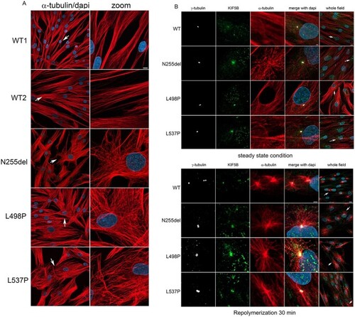
Defective KIF5B function impacts MT orientation and cell shape and localize to MTOC during MT repolymerization as well as in interphase. (A) Confocal microscopy analyses show an evident aberrant orientation of MT in patients’ cells in interphase compared to control cells. Scale bar is 5 μm (B) Panel show a colocalization of KIF5B with the MTOC. In patients’ cells this colocalization is evident during MT repolymerization but persist during cell interphase. In WT cells, it is more evident during MT repolymerization and less in cells in steady state condition. This experiment suggests a role of KIF5B on microtubular dynamics. Scale bar is 2 μm (zoomed images) and 10 μm (whole field) respectively. Cells were stained with anti-α-tubulin antibody (MT marker, red), anti-γ-tubulin (MTOC marker, white), KIF5B (green) and DAPI (blue). |