
Fig. 1
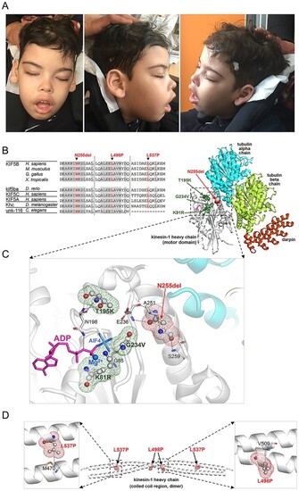
Clinical features of subject 1 and structural data. (A) The subject (8 years) showed hypotonic face, bitemporal narrowing, bushy and straight eyebrow, hypertelorism, long eyelashes, long palpebral fissures, large ears with thick lobe, broad nasal bridge, anteverted nares, short deep philtrum, everted upper lip and prominent upper and lower vermilion. Explicit permission was obtained to publish photographs of the subject. (B) Multiple sequence alignment of KIF5B orthologs and paralogs around the mutated residues. Residues that are invariant (at least down to Drosophila melanogaster) are grayed. (C) The 3D structure of the human kinesin-1 heavy chain motor domain complexed with tubulin and darpin (PDB 4HNA) showing Asn255 (balls and sticks with red meshes), its interactions (dotted lines) with nearby residues (sticks), the co-crystallized ADP (magenta sticks), and Mg2+ cation (blue). The residues mutated in KIF5B-related KD (Lys91, Thr195, and Gly234; balls and sticks with green meshes) are also shown. The pathogenic missense changes are predicted to perturb KIF5B interaction with ATP/ADP. (D) 3D structure of the coiled-coil region (dimeric) of human kinesin-1 heavy chain (PDB 6IGV) including the site affected by the p.Leu498Pro e p.Leu537Pro substitutions (displayed on both monomers). Leu498 in one monomer interacts with Val509 on the bound monomer, while Leu537 on one monomer interacts with Met470 on the bound monomer. |