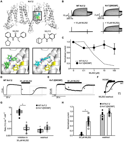
Kv7.2 Trp236 strongly influences ML252 inhibition. (A) Docked poses of ML252 (green) in the cryo-EM structure of Kv7.2 (PDB 7VNP), along with the experimentally reported binding mode of ML213 (cyan). The binding pocket is enlarged in the inset, illustrating the orientation of ML252 (left) or ML213 (right) relative to Trp236 (yellow). (B) Exemplar traces from Xenopus oocytes expressing WT Kv7.2 (left) or Kv7.2[W236F] (right), in control or 10 μm ML252, as indicated. Voltage was held at −80 mV and pulsed between −140 and +40 mV (10 mV steps). (C) ML252 concentration response of Kv7.2[W236F] in Xenopus oocytes at +20 mV (n = 5) (WT Kv7.2 data from Figure 1F re-plotted as dashed line). (D and E) Exemplar patch-clamp current recordings of (D) WT Kv7.2 or (E) Kv7.2[W236F] expressed in HEK293 cells. Cells were depolarized and held at +20 mV, followed by drug application or washout where indicated. Segments a and b were used to measure the rate of onset of inhibition, segments c and d were used to measure the rate of recovery. (F) Expanded view of the kinetics of current recovery after ML252 washout in WT Kv7.2 or Kv7.2[W236F] (boxes c and d from panels D and E). (F and G) Traces obtained from ML252 washouts expressing WT Kv7.2 were fit with a Hodgkin–Huxley equation and Kv7.2[W236F] traces were fit with a single exponential function. (G) Rates of ML252 inhibition and recovery in WT Kv7.2 (recovery n = 11, inhibition n = 20) or Kv7.2[W236F] (recovery n = 17, inhibition n = 28) (* indicates P < .001, Student’s t-test). (H) Current magnitudes (normalized to maximal current at +20 mV) in ML252 or after washout, for WT Kv7.2 (n = 12 in 30 μm ML252, n = 8 washout) or Kv7.2[W236F] (n = 25 in 30 μm ML252, n = 15 washout), (* indicates P < .001, Student’s t-test of current inhibition of WT 7.2 vs. 7.2[W236F]).
|
