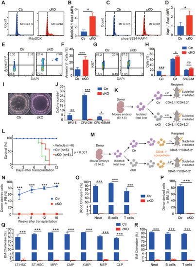
Slc39a10‐deficient HSCs have increased ROS levels, increased DNA damage, a higher prevalence of apoptosis, and impaired repopulation capacity. A,B) Representative FACS profiles (A) and quantification (B) of MitoSOX mean fluorescence intensity (MFI) measured in HSCs (Lin−Sca1+Mac1+) obtained from control and cKO embryos at E14.5 (n = 3 per group). C,D) Representative FACS profiles (C) and quantification (D) of intracellular p‐Kap1 staining in HSCs (Lin−Sca1+Mac1+) obtained from control and cKO embryos at E14.5 (n = 4 and 3 for control and cKO, respectively). E,F) Representative FACS profiles (E) and quantification (F) of annexin V staining in LT‐HSCs (Lin−Sca1+CD48−CD150+Mac1+) obtained from control and cKO embryos at E14.5 (n = 4 per group). G,H) Representative FACS profiles of intracellular Ki67 staining in HSCs (Lin−Sca1+Mac1+) obtained from control and cKO embryos at E14.5 (G), and summary of the percentage of cells in the indicated cell cycles (H; n = 3 per group). I,J) Representative images of in vitro colony assays performed using fetal liver cells obtained from control and cKO embryos at E14.5 (I), and summary of the number of primitive erythroid progenitor cells (BFU‐E), granulocyte‐macrophage progenitor cells (CFU‐GM), and multipotent granulocytes, erythroid, macrophage, and megakaryocyte progenitor cells (CFU‐GEMM) measured on day 12 (n = 3 per group). K) Schematic diagram depicting the transplantation strategy in which lethally irradiated CD45.1+/CD45.2+ recipient mice received donor‐derived (CD45.2+) fetal liver cells. L) Kaplan–Meier survival curve of CD45.1+/45.2+ mice that were lethally irradiated and then transplanted with vehicle or fetal liver cells obtained from either CD45.2+ control or cKO embryos (n = 6 recipients per group). M) Schematic diagram depicting the strategy for competitive transplantation in which lethally irradiated CD45.1+/CD45.2+ recipient mice received the indicated donor‐derived fetal liver (CD45.2+) cells and competitor (CD45.1+) cells at a 1:1 ratio. N) Time course showing the percentage of donor‐derived CD45.2+ cells in the peripheral blood of recipient mice measured at the indicated time points following transplantation (n = 5 recipients per group). O) Summary of the percentage of donor‐derived myeloid cells, B cells, and T cells in the peripheral blood of recipient mice 16 weeks after co‐transplantation with control or cKO cells (n = 5 recipients per group). P) Summary of the percentage of donor‐derived cells (CD45.2+) in the bone marrow of recipient mouse mice 16 weeks after transplantation (n = 5 recipients per group). Q) Summary of the percentage of donor‐derived LT‐HSCs, short‐term HSCs (ST‐HSCs), MPP (multipotent progenitor cells), CMP (common myeloid progenitor cells), GMP (granulocyte/monocyte progenitor cells), MEP (megakaryocyte/erythrocyte progenitor cells), and CLP (common lymphoid progenitor cells) 16 weeks after co‐transplantation with control or cKO donor cells (n = 5 recipients per group). R) Summary of the percentage of donor‐derived myeloid cells, B cells, and T cells in the bone marrow of recipient mice 16 weeks after co‐transplantation with control or cKO cells (n = 5 recipients per group). Data in this figure are represented as mean ± SD. p values of survival in (L) were determined using the Log‐rank test, in (B), (D), (F), (H), (J), (N), (O), (P), (Q), and (R) using 2‐tailed unpaired Student's t‐test. *p < 0.05, ***p < 0.001, and ns, not significant.
|
