
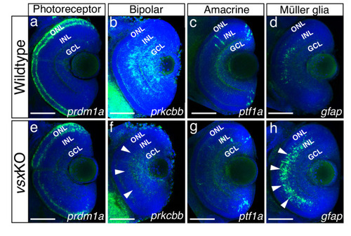
Figure 4—figure supplement 1.
- ID
- ZDB-FIG-230526-5
- Publication
- Letelier et al., 2023 - Mutation of vsx genes in zebrafish highlights the robustness of the retinal specification network
- Other Figures
-
- Figure 1—figure supplement 1.
- Figure 1—figure supplement 1.
- Figure 1—figure supplement 2.
- Figure 1—figure supplement 3.
- Figure 2—figure supplement 1.
- Figure 2—figure supplement 1.
- Figure 3—figure supplement 1.
- Figure 3—figure supplement 1.
- Figure 4—figure supplement 1.
- Figure 4—figure supplement 1.
- Figure 4—figure supplement 2.
- Figure 4—figure supplement 3.
- Figure 5—figure supplement 1.
- Figure 5—figure supplement 1.
- Figure 5—figure supplement 2.
- Figure 5—figure supplement 3.
- Figure 5—figure supplement 4.
- All Figure Page
- Back to All Figure Page
(a-f), (h-m). Cryosections of wildtype (a-c, h-j) and vsxKO retinas (d-f, k-m) stained with photoreceptor specific antibodies and DAPI as a nuclear marker. Cones and rods were visualized by using the Zpr1 (a-f) and Zpr3 (h-m) antibodies, respectively. Delayed cone and rod differentiation is observed in vsxKO mutants (d, k) compared to wildtype (a, h) samples at 72hpf (white arrowheads in d, k). At 96hpf, both markers are expressed in similar levels in wildtype (b, i) and double mutant retinas (e, l), but at 6dpf there is a significant increase of Zpr1 fluorescent intensity in vsxKO (f) compared to WT retinas (c) using an unpaired t-test (*p<0.05). No major differences were observed in rod stain intensity between WT (j) and vsxKO (m) at 6dpf (p>0.05). (g, n). Quantification of cones (g) and rods (n) label intensity between WT and vsxKO retinas were performed using Fiji software. Fluorescent measurements in the ONL were normalized by background signal from the INL. Data is shown as mean ± SD. For all conditions tested (except for c, f, j) n≥10. ONL: outer nuclear layer, INL: inner nuclear layer, GCL: ganglion cell layer, h: hours post-fertilization, d: days post-fertilization, ns: not significant. |