
Figure 1—figure supplement 3.
- ID
- ZDB-FIG-230526-4
- Publication
- Letelier et al., 2023 - Mutation of vsx genes in zebrafish highlights the robustness of the retinal specification network
- Other Figures
-
- Figure 1—figure supplement 1.
- Figure 1—figure supplement 1.
- Figure 1—figure supplement 2.
- Figure 1—figure supplement 3.
- Figure 2—figure supplement 1.
- Figure 2—figure supplement 1.
- Figure 3—figure supplement 1.
- Figure 3—figure supplement 1.
- Figure 4—figure supplement 1.
- Figure 4—figure supplement 1.
- Figure 4—figure supplement 2.
- Figure 4—figure supplement 3.
- Figure 5—figure supplement 1.
- Figure 5—figure supplement 1.
- Figure 5—figure supplement 2.
- Figure 5—figure supplement 3.
- Figure 5—figure supplement 4.
- All Figure Page
- Back to All Figure Page
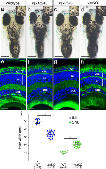
(a-d). Dorsal view of wildtype (a), vsx1∆245 (b), vsx2∆73 (c), and vsxKO (d) animals at 6dpf. vsx1∆245 (b) and vsxKO (d) fish have problems sensing background light and appear darker than wildtype (a) and vsx2∆73 (c) larvae, as melanosomes are broadly distributed within melanophores (white arrowheads). (e-h). Histological sections of wildtype (e), vsx1∆245 (f), vsx2∆73 (g), and vsxKO (h) eyes at 6dpf. In the central retina of vsxKO double mutants (h), the INL width is reduced and the ONL thickness is expanded compared to wildtype (e), vsx1∆245 (f), or vsx2∆73 (g) samples. DAPI and phalloidin coupled to Alexa488 were used as nuclear and cytoskeletal (actin) markers, respectively. (i). Quantification of INL (blue circles) and ONL (green squares) width in central retinas at 6dpf using histological sections as in (e-h) panels. Significant reduction and expansion of the INL and ONL, respectively, was observed in vsxKO retinas when compared to wildtype (***p<0.0001) using an unpaired t-test. Data are shown as mean ± SD. ONL: outer nuclear layer, INL: inner nuclear layer, GCL: ganglion cell layer, µm: micrometres. Scale bar in (a-d): 500µm, scale bar in (e-h): 20µm. |