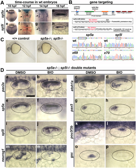
Early expression of sp5a and sp5l and effects of disruption on otic patterning. (A) Dorsal views with anterior to the top (Aa-c, Ae-Ag) and dorsal views with anterior to the left (Ad, Ah) showing expression of sp5a and sp5l in wild type embryos at the times indicated at the top. Co-staining for krox20 (red) is shown at 12 hpf (Ab, Af) to mark positions of rhombomeres 3 and 5 (r3, r5) in the hindbrain. The midbrain-hindbrain border (mhb) is labeled. Arrows point to otic domains (Ab, Ac, Af, Ag). (B) Targeting of sp5a and sp5l. The upper diagram represents the general layout of both genes, including regions encoding specific peptide domains. Red arrows show relative positions of deletions induced by CRISPR/Cas9 cutting. Altered sequences in mutant alleles sp5ax69 and sp5lx70 are shown in boxed regions and in sequencing traces at the bottom. (C) Lateral views of live wild-type and sp5a−/−; sp5l−/− embryos at 24 hpf. (D) Dorsolateral views of sp5a−/−; sp5l−/− double mutants showing expression at 24 hpf of various otic genes (as indicated on the left) following treatment with DMSO or BIO from 12 hpf (as indicated at the top). Scale bar, 50 μm.
|
