
Figure 3
- ID
- ZDB-FIG-230228-13
- Publication
- Enders et al., 2023 - Impact of the Voltage-Gated Calcium Channel Antagonist Nimodipine on the Development of Oligodendrocyte Precursor Cells
- Other Figures
- All Figure Page
- Back to All Figure Page
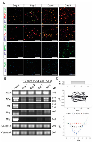
Characterization of primary OPCs after stimulation with PDGF and FGF-2. (A) Immunocytochemistry of OPCs with MBP (red)/Olig2 (green), PLP (red)/SOX10 (green), MAG (green), and MOG (green) after 1, 2, 4, and 6 days, respectively, following stimulation with 10 ng/mL PDGF and FGF-2. Scale bars represent 100 µm. (B) End-point PCR after stimulation with 10 ng/mL PDGF and FGF-2. Values on the right indicate the length of the PCR products in base pairs. (C) Patch-clamp experiments revealed the physiological presence of VGCCs, which could be blocked by 10 µM nimodipine and activated by 10 µM BayK8644. DMSO was used as control agent. Data are presented as mean ± standard deviation. All experiments were performed as n = 3 independent experiments. Actb: beta-actin; BayK: BayK8644; bp: base pairs; Cacna1c: voltage-gated L-type calcium channel 1.2; Cacna1d: voltage-gated L-type calcium channel 1.3; DAPI: 4′,6-diamidino-2-phenylindole; DMSO: dimethyl sulfoxide; FGF-2: fibroblast growth factor 2; Mag: myelin-associated glycoprotein; Mbp: myelin basic protein; Mog: myelin oligodendrocyte glycoprotein; ms: milliseconds; mV: millivolt; Nimo: nimodipine; pA: picoamperes; PDGF: platelet-derived growth factor; Plp: proteolipid protein; SOX10: SRY-box transcription factor 10; VGCC: voltage-gated L-type calcium channel. |