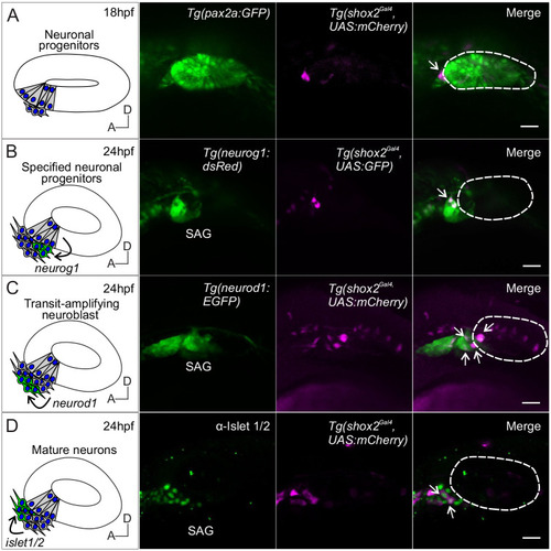
Lineage labeling of neurogenic cells in the developing SAG using shox2 fluorescent reporter. Fluorescent images of developing inner ear from Tg(shox2Gal4,UAS:GFP) reporter larva with inner ear and neuronal reporters. Curved arrows on diagrams indicate migration of developing neurons from its previous locations. Dashed lines in merged images outline the otic vesicle. (A) Diagram of neuronal progenitors in the anteroventral region of the otic vesicle at 18 hpf. Reporter fluorescence from a pan-otic Tg(pax2a: GFP) and Tg (shox2Gal4,UAS:mCherry) at 18 hpf. Merged image depicts shox2 reporter labeled cells (arrow) in the anteroventral region adjacent to the otic vesicle (n= 5). (B) Diagram of delaminating neuroblasts expressing neurog1 (green) at the floor of the otic vesicle. Fluorescence from Tg(neurog1: dsRed) and Tg(shox2Gal4,UAS:GFP) reporters at 24 hpf. Merged image identifies specified neuroblasts and delaminating neuroblasts (arrows) (n=14 embryos). (C) Diagram of transit-amplifying neuroblasts expressing neurod1 (green) at 24 hpf after delaminating from the otic vesicle. Fluorescence from TgBAC(neurod1: EGFP) and Tg(shox2Gal4,UAS:mCherry) reporters at 24 hpf. Merged image shows overlap of neurod1 and shox2 reporter fluorescence (arrows) (n=9 embryos). (D) Diagram of mature neurons expressing Islet 1/2 (green) from transit amplifying neuroblasts (arrow). Fluorescence from the medial position of the neurogenic domain shows Islet 1/2 immunostaining and Tg(shox2Gal4, UAS:mCherry) reporter at 24 hpf. Merged image shows overlap of Islet 1/2 and shox2 reporter marked cells (arrow) (n= 11 embryos). SAG, statoacoustic ganglion. Scale bars: 10 µm.
|
