
Figure 3
- ID
- ZDB-FIG-221029-24
- Publication
- Davison et al., 2022 - Zebrafish Slit2 and Slit3 Act Together to Regulate Retinal Axon Crossing at the Midline
- Other Figures
- All Figure Page
- Back to All Figure Page
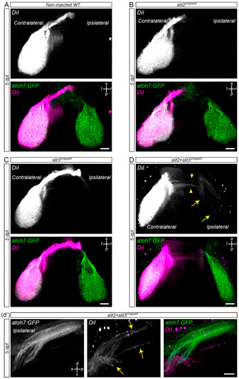
Retinal axons from slit2 + slit3 crispants innervate the ipsilateral optic tectum. Retinotopic anterograde RGC axon DiI labeling in atoh7:Gap43-EGFP (atoh7:GFP) transgenic larvae. (A–D). Horizontal maximum intensity z-projections of the cephalic region of 5 dpf larvae, where both the contralateral and ipsilateral tecta can be observed (dorsal view). Non-injected wild-type (A), slit2 crispant (B), slit3 crispant (C) and slit2 + slit3 crispant (D) larvae are shown. DiI-labeled axons are observed in the ipsilateral tectum of slit2 + slit3 crispants (arrows), and can be better visualized in the rotated view in d’. DiI-labeled axons can also be observed crossing the midline at a site slightly posterior to the optic tract (arrowheads). Scale bars: (A–D), 30 μm; (d’), 20 μm. See Video S3. |