
Fig. 3
- ID
- ZDB-FIG-220927-67
- Publication
- Zou et al., 2022 - Macrophages Rapidly Seal off the Punctured Zebrafish Larval Brain through a Vital Honeycomb Network Structure
- Other Figures
- All Figure Page
- Back to All Figure Page
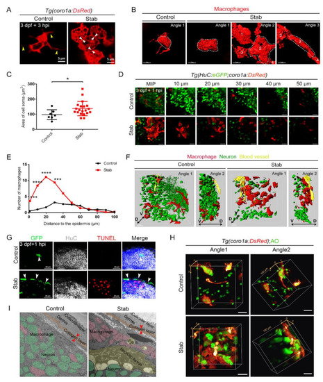
Aggregated macrophages formed a honeycomb network structure covering the wound surface. (A) Changes in macrophages morphology after injury. The yellow arrowheads indicate the branches. The white arrowheads indicate tight connections between cells. Scale bar, 5 µm. (B) Three-dimensional reconstruction of the morphology of macrophages after injury. Angle 1 shows the morphology of individual cells and Angles 2 and 3 indicate tight connections between cells. Scale bar, 7 µm. (C) Statistic analysis of the area of cell soma surface at 3 hpi. Control, 97.08 ± 12.37 µm2 n = 7; Stab, 137.50 ± 10.61 µm2 n = 20. (D) Distribution of macrophages and neurons on different layers of the Z axis at 1 hpi. The number on each panel represents the distances to the surface. Scale bar, 20 µm. (E) Statistics analysis of coro1a+ cells number at different layers with distinct distances from the epidermis at 1 hpi. (F) Three-dimensional images of the distribution of macrophages (red) and neuron (green) in Tg(coro1a:DsRed;HuC:eGFP) at 1 hpi. Yellow marks the blood vessels. D, dorsal; V, ventral. Scale bar, 15 µm. (G) The immunofluorescent images of GFP, HuC and TUNEL on the frozen sections of Tg(coro1a:eGFP) brains at 1 hpi. The white arrowheads present the distribution of GFP+ cells. Scale bar, 20 µm. (H) The location of macrophages and AO+ signals in 3D view. Scale bar, 20 µm. (I) Transmission electron microscope image. Macrophages are in pink, neurons are in green, apoptotic or necrotic tissue is in yellow and the collagen layer beneath the skin is in orange. The red arrowheads indicate the width of the collagen layer. Scale bar, 5 µm. (Data are shown as mean ± SEM. *, p < 0.05; ***, p < 0.001; ****, p < 0.0001.) |