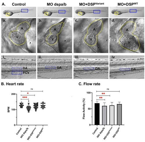
Zebrafish DSP Model Cardiac examination at 72 hours post fertilization (hpf). Modeling Qatar Genome Programme human DSP cardiac variant produced specific cardiac phenotype in the zebrafish model, representative heart images at 72 hours post-fertilization (hpf): (A) Zebrafish larvae at 72 hpf showing the two chambered heart (yellow square) and dorsal aorta flow rate (blue rectangle) images at 32X magnification. Representative images of the heart chambers; atrium (a) and ventricle (v) (traced with yellow dotted line) in the zebrafish experimental groups (control, Morpholino injected (MO dspa/b), human synthetic RNA of variant DSP c.1841A > G (p.Asp614Gly) (MO + DSPVariant) and wild-type (MO + DSPWT) co-injected with corresponding MO targeting the endogenous zebrafish transcript). (B) Heart rate for each individual fish was calculated as beats per minute (bpm) for the four experimental groups. Each dot represents an animal. Average heart rate of each group was compared to the control group. (C) The analysis of the vascular parameters was performed to calculate the blood flow rate within a selected area of the blood vessel (dorsal aorta). Average blood flow activity of each group was compared to the control group. Control (n = 25), dspa/b MO (n = 28), MO + DSPVariant (n = 46), and MO + DSPWT (n = 24). ns: not significant.
|
