
FIGURE
Fig. 6
- ID
- ZDB-FIG-220809-48
- Publication
- Yáñez et al., 2021 - The organization of the zebrafish pallium from a hodological perspective
- Other Figures
- All Figure Page
- Back to All Figure Page
Fig. 6
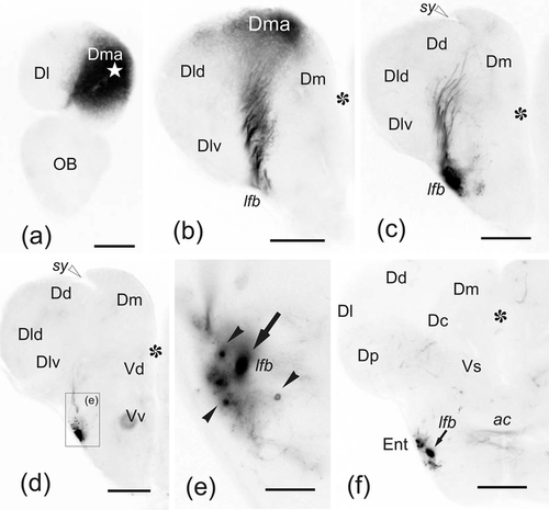
(a–f) Photomicrographs of transverse sections through the telencephalic lobes of zebrafish, from rostral to caudal, showing labeled cell bodies (black arrowheads) and fibers (arrows) after application of DiI to the rostralmost medial zone of the pallium (Dma). (a) Labeling of Dma around the accessed region (white star). (b–c) Sections of more caudal regions showing scant Dma connections with adjacent pallial regions (b), and small fiber bundles coursing toward the (lfb) through deeper pallial regions. (d–f) A terminal field labeled in the entopeduncular nucleus. In (e), note afferent neurons to Dma (arrowheads). Asterisk: telencephalic ventricle. Midline is to the right. Ipsilateral side is to the left. (e) is a detail of the squared area in (d). For abbreviations, see the list. All photomicrographs are negative images of fluorescent data. White arrowheads: sulcus ypsiloniformis. Scale bars: 200 μm (a–d, f); 50 μm (e)
|
Expression Data
Expression Detail
Antibody Labeling
Phenotype Data
Phenotype Detail
Acknowledgments
This image is the copyrighted work of the attributed author or publisher, and
ZFIN has permission only to display this image to its users.
Additional permissions should be obtained from the applicable author or publisher of the image.
Full text @ J. Comp. Neurol.