
FIGURE
Figure 2
- ID
- ZDB-FIG-220430-81
- Publication
- Lin et al., 2022 - Cerebroventricular Injection of Pgk1 Attenuates MPTP-Induced Neuronal Toxicity in Dopaminergic Cells in Zebrafish Brain in a Glycolysis-Independent Manner
- Other Figures
- All Figure Page
- Back to All Figure Page
Figure 2
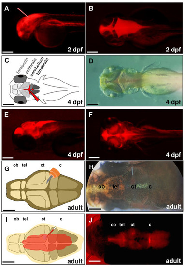
Figure 2. Distribution of the injected exogenous DsRed protein in brain chamber. We injected exogenous red fluorescence protein (DsRed) into the brain chamber of a zebrafish embryo at (A,B) 2-, (C–F) 4-dpf, and (G–J) a 6-month-old adult zebrafish. (A) The glass needle represents the injection site of zebrafish embryo’s head at 2 dpf. (C) Schematic diagram depicts the direct injection of DsRed into the brain chamber of zebrafish embryo at 4 dpf. The glass needle marked in red represents the injection site. (D) Dorsal view of 4-dpf embryo injected with DsRed under bright-field microscopy; (E) Lateral view of 4-dpf embryo injected DsRed under fluorescence microscopy; (F) Dorsal view of 4-dpf embryo injected DsRed under fluorescence microscopy. (G) Schematic diagram depicts a small incision that was generated above the optic tectum of adult zebrafish by a 30-gauge syringe. (H) Dorsal view of adult zebrafish injected DsRed by glass needle under bright-field microscopy; (I) Schematic diagram depicts the distribution of injected DsRed in the brain through the cerebroventricular fluid. (J) Dorsal view of adult zebrafish injected DsRed after 30 min under fluorescence microscopy. ob: olfactory bulb, tel: telencephalon, ot: optic tectum, c: cerebellum. Scale bar: (A–F) 25 µm; (G–J) 500 µm.
|
Expression Data
Expression Detail
Antibody Labeling
Phenotype Data
Phenotype Detail
Acknowledgments
This image is the copyrighted work of the attributed author or publisher, and
ZFIN has permission only to display this image to its users.
Additional permissions should be obtained from the applicable author or publisher of the image.
Full text @ Int. J. Mol. Sci.