FIGURE

FIGURE 6

ID
ZDB-FIG-220420-41
Publication
Martins et al., 2022 - Müller Glia maintain their regenerative potential despite degeneration in the aged zebrafish retina
Other Figures
All Figure Page
Back to All Figure Page
FIGURE 6

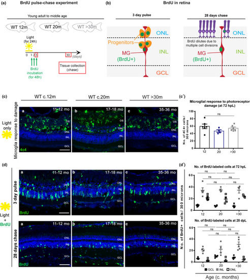
Zebrafish MG regenerative capacity upon acute damage is maintained in old age. (a) Schematic image of the experimental design and (b) expected results. Fish were light‐treated for 24 h and BrdU incorporation occurred between 24–72 h (green), which allowed for BrdU to be washed out the proliferating progenitors, leaving only MG which re‐entered the cell cycle to be labelled. (c) The central retina labelled with 4C4 (microglia, in green), after light treatment, in adults (c. 12 months), middle aged (c. 20 months) and old (>30 months) albino zebrafish. The majority of the microglia response to damage occurs within the OS and ONL of the retina, where a debris field is present due to photoreceptor degeneration. (c’) Quantification of the number of microglial cells which responded to photoreceptor damage in the different aged groups. (d, top) The central retina 72 h after light treatment onset, immunolabelled with BrdU (proliferation, in green). (d’). Quantification of the number of cells proliferating in the GCL (represented in black squares), INL (represented in grey squares), and ONL (represented in white squares) in each age group. (d, bottom) The central retina 28 days after light treatment onset. (d’). Quantification of the number of BrdU‐positive cells observed in the GCL, INL, and ONL of each aged group. Error bars represent the SEM. N = 4–5. Scale bars: 20 μm

Expression Data

Expression Detail
Antibody Labeling
Phenotype Data

Phenotype Detail
Acknowledgments
This image is the copyrighted work of the attributed author or publisher, and ZFIN has permission only to display this image to its users. Additional permissions should be obtained from the applicable author or publisher of the image. Full text @ Aging Cell