FIGURE

FIGURE 2

ID
ZDB-FIG-220420-37
Publication
Martins et al., 2022 - Müller Glia maintain their regenerative potential despite degeneration in the aged zebrafish retina
Other Figures
All Figure Page
Back to All Figure Page
FIGURE 2

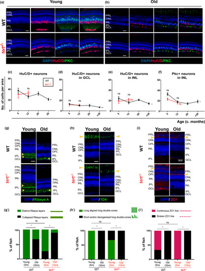
Zebrafish retina undergoes neurodegeneration with ageing, independently of telomerase. The central retina immunolabelled with HuC/D and PKC (amacrine in magenta and bipolar cells in green, respectively), in both WT and tert−/−, in (a) young (5 months) and (b) old adults (>30 months in WT and 12 months in tert−/−). Scale bars: 20 μm. (c–f) Quantifications of the number of HuC/D‐positive neurons per area (10,000 μm2) (c) in the overall retina, and (d) in the GCL (ganglion cells) and (e) INL (amacrine cells). (f) Quantifications of the number of PKC‐positive neurons cells per area (10,000 μm2) in the INL (bipolar cells). Error bars represent the SEM. N = 3–7. (g–i) The central retina immunolabelled with (g) Ribeye A (BC synaptic terminals, in green), (h) 1D4 (outer segments of long double‐cones, in green), and (i) ZO1 (outer limiting membrane, in red), in both WT and tert−/−, in young (5 months) and old adults (>30 months in WT and 12 months in tert−/−). Scale bars: 20 μm. (g’–i’) Quantification of the percentage of fish presenting defects in (g’) Ribeye A (phenotype observed as disorganised synaptic terminal layering in the IPL), (h’) 1D4 (short and/or misaligned long double‐cone outer segments), and (i’) ZO1 (broken outer limiting membrane). Error bars represent the SEM. (g’) N = 4–9, (h’, i’) N = 3–6

Expression Data

Expression Detail
Antibody Labeling
Phenotype Data

Phenotype Detail
Acknowledgments
This image is the copyrighted work of the attributed author or publisher, and ZFIN has permission only to display this image to its users. Additional permissions should be obtained from the applicable author or publisher of the image. Full text @ Aging Cell