FIGURE

FIGURE 4

ID
ZDB-FIG-220420-39
Publication
Martins et al., 2022 - Müller Glia maintain their regenerative potential despite degeneration in the aged zebrafish retina
Other Figures
All Figure Page
Back to All Figure Page
FIGURE 4

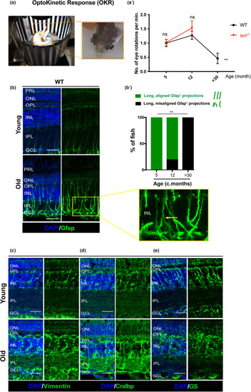
Zebrafish vision declines with ageing, which is accompanied by signs of glial morphological alterations. (a) OKR assay was performed by immobilising the fish in between soft sponges, inside a petri dish containing water, placed in the centre of a rotation chamber. The walls of the rotation chamber had 0.8 mm‐thick black and white stripes and the chamber was maintained at a constant velocity of 12 rpm throughout the experiment. (a’) The number of eye rotations per minute was manually quantified by video observation. Error bars represent the SEM. N = 5–8. (b) MG display signs of altered morphology at old ages, as evidenced by Gfap staining. The central retina immunolabelled with Gfap (in green), in WT, young (5 months) and old adults (>30 months). Yellow arrows highlight an example of aligned versus misaligned MG projections. (b’) Quantifications of the percentage of fish presenting disorganised MG processes in the IPL (detailed view‐inset). N = 6. (c–e) MG in the old retina show altered morphology and altered expression of stress proteins. The central retina immunolabelled with (c) Vimentin, (d) Cralbp, and (e) GS showing disorganised MG in old adults (>30 months) compared with young adults (5 months). N = 6. Scale bars: 20 μm

Expression Data

Expression Detail
Antibody Labeling
Phenotype Data

Phenotype Detail
Acknowledgments
This image is the copyrighted work of the attributed author or publisher, and ZFIN has permission only to display this image to its users. Additional permissions should be obtained from the applicable author or publisher of the image. Full text @ Aging Cell