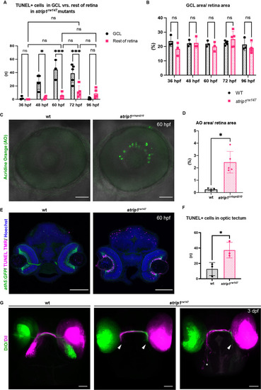
<italic toggle='yes'>strip1</italic> mutants show apoptosis in retinal ganglion cells (RGCs) and optic tectum, and elongation defects in retinal axons.
(A) The number of transferase dUTP nick end labeling (TUNEL+) in the ganglion cell layer (GCL) compared to the rest of the retina at 36, 48, 60, 72, and 96 hpf in strip1rw147 mutant retinas, for experiments shown in Figure 3A. Apoptosis gradually increases to reach its peak at 72 hpf in other retinal areas, but its number at 72 hpf is significantly lower than that of the GCL at 72 hpf. In addition, there is no significant difference in apoptotic cell number of other retinal areas between 72 hpf and other time points (36, 60, and 96 hpf). Two-way analysis of variance (ANOVA) with the Tukey multiple comparison test, n ≥ 3. (B) Percentage of GCL area (area between the lens and inner plexiform layer [IPL]) relative to the total retinal area in wild-type and strip1rw147 mutant retinas at 36, 48, 60, 72, and 96 hpf, for experiments shown in Figure 3A. There is no significant difference between wild-type siblings and strip1rw147 mutants at any stage. Two-way ANOVA with the Tukey multiple comparison test, n ≥ 3. (C) Live confocal images of wild-type and strip1crisprΔ10 mutant retinas stained with acridine orange (AO) to label apoptotic cells at 60 hpf. Scale bar, 50 μm. (D) Percentage of AO area relative to total retina area in 60 hpf wild-type and strip1crisprΔ10 mutant retinas. Mann–Whitney U-test, n = 5. (E) TUNEL of 60 hpf wild-type and strip1rw147 mutant heads combined with Tg[ath5:GFP] to label RGCs. All nuclei are counterstained with Hoechst. Apoptosis occurs markedly in RGCs and optic tectum in strip1rw147 mutants. Scale bar, 100 μm. (F) The number of TUNEL+ in the optic tectum of wild-type and strip1rw147 mutants at 60 hpf. The number of TUNEL+ cells is significantly higher in strip1rw147 mutants than in wild-type siblings. Student’s t-test with Welch’s correction, n = 3. (G) Dorsal view of the optic tectum of 3-dpf wild-type and strip1rw147 mutants. RGC axons are labeled using intraretinal injections of DiO or DiI. Axons of strip1rw147 appear to exit the optic disc normally and the optic chiasm is formed. However, the optic nerve is thinner in strip1rw147 mutants than in wild-type siblings. In addition, most axons fail to elongate properly and do not arborize within the optic tectum (arrowheads) or very few axons elongate poorly to reach more posterior arborization fields within the optic tectum (asterisk). Scale bar, 50 μm. For all graphs, data are represented as means ± standard deviation (SD). ns, not significant, *p < 0.05, and ***p < 0.001.
Data for <xref rid='fig3s1' ref-type='fig'>Figure 3—figure supplement 1A,B,D,F</xref>.
Expression Data
Expression Detail
Antibody Labeling
Phenotype Data
Phenotype Detail
Acknowledgments
This image is the copyrighted work of the attributed author or publisher, and
ZFIN has permission only to display this image to its users.
Additional permissions should be obtained from the applicable author or publisher of the image.
Full text @ Elife
Your Input Welcome
Thank you for submitting comments. Your input has been emailed to ZFIN curators who may contact you if
additional information is required.
Oops. Something went wrong. Please try again later.