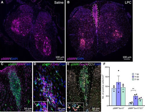
Expression of pS6RP in LPC-induced demyelinated lesions. (A) Expression of pS6RP in the saline-injected spinal cord. (B) pS6RP immunolabeling in LPC-induced demyelinated spinal cord at 7 dpi. pS6RP is upregulated in the LPC lesion of the dorsal funiculus (dashed lines). (C) Co-immunolabelling of pS6RP and GFAP. Note that GFAP+ astrocytes are not stained with the anti-pS6RP antibody. (D) Co-immunolabelling of pS6RP and CD45 reveals expression in few monocytes/macrophages (arrowhead, inset). (E) Triple immunostaining for pS6RP, Sox10 and CC1. Inset (E) illustrates the expression of pS6RP in Sox10+CC1+ oligodendrocytes in the lesion at 7 dpi (arrowheads). (F) Quantification of pS6RP-expressing oligodendrocytes showed a peak of pS6RP expression at 14 dpi, coinciding with the remyelination phase of the lesion. Data represent mean ± SEM; N = 3 independent experiments at each timepoint. ANOVA followed with post hoc Tukey's pairwise multiple comparison tests: **P ≤ 0.01. Scale bars: (A and B), 100 µm; (C and E), 50 µm; (D), 20 µm.
|
