|
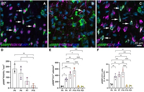
Characterization of pS6RP expression in oligodendroglial cells. (A–C) Co-immunolabelling for pS6RP and Sox10 (A), pS6RP and PDGFRα (B), pS6RP and CC1 (C) in the dorsal funiculus of PND7 WT mice. Double-positive cells are indicated by arrows. (D) Number of pS6RP+PDGRα+ OPCs/mm2 from PND0 to PND10. (E) The number of CC1+ differentiated oligodendrocytes expressing pS6RP from PND0 to PND21. Note that pS6RP is transiently expressed in CC1+ differentiated oligodendrocytes. (F) Although transient, pS6RP is predominantly located in CC1+ differentiated oligodendrocytes between PND4 and PND10. Data represent mean ± SEM; N = 3 independent experiments for each developmental stage. ANOVA followed with post hoc Tukey's pairwise multiple comparison tests: *P < 0.05; **P < 0.01; ***P ≤ 0.001; ****P ≤ 0.0001. Panels (A–C) are counterstained with DAPI. Scale bar (A–C): 20 µm.
|