
Figure 4
- ID
- ZDB-FIG-220302-83
- Publication
- Vagionitis et al., 2022 - Clusters of neuronal neurofascin prefigure the position of a subset of nodes of Ranvier along individual central nervous system axons in vivo
- Other Figures
- All Figure Page
- Back to All Figure Page
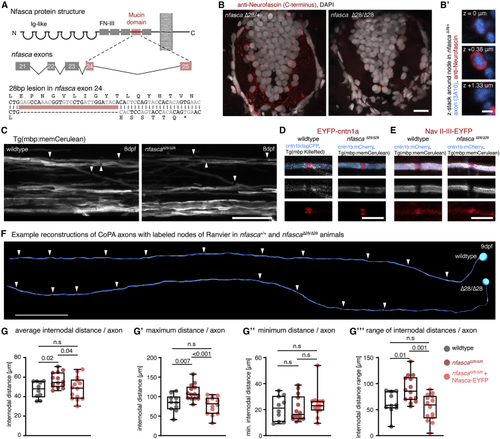
Increased and less regular nodal spacing along individual axons in (A) Targeting strategy and genetic lesion in (B) Immunohistochemistry against Nfasca on spinal cord cross-sections in adult heterozygous and homozygous (C) Overviews of the spinal cord in full transgenic myelin reporter lines. Arrowheads highlight gaps between adjacent myelin sheaths. Scale bar, 20 μm. (D and E) Close-up views showing single nodes of Ranvier marked with EYFP-cntn1a and EYFP-NaV-II-III in full transgenic myelin reporter lines. Scale bar, 5 μm. (F) Reconstructions of two CoPA neurons and their node of Ranvier positions (arrowheads) from wild type and (G) Average internodal distances along single axons per axon in wild type, (G′–G‴) Maximum (G′), minimum (G″), and range (G‴) of internodal distances per axon as quantified in (G). Median with IQR; Brown-Forsythe ANOVA with multiple comparisons for (G′ and G‴), Kruskal-Wallis test with multiple comparisons for (G″). See also |
| Gene: | |
|---|---|
| Antibody: | |
| Fish: | |
| Anatomical Terms: | |
| Stage Range: | Day 4 to Days 7-13 |
| Fish: | |
|---|---|
| Observed In: | |
| Stage Range: | Day 4 to Days 7-13 |