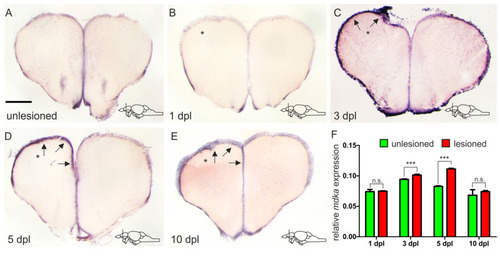
mdka is upregulated in a delayed fashion in response to injury. (A–E) ISH against mdka on cross-sections of the adult telencephalon in an unlesioned brain (A) and at different time points after the lesion (B–E). (B) Immediately after inflicting the lesion, there is no upregulation of mdka expression in the injured hemisphere. (C–E) mdka expression is increased in the injured hemisphere from 3 dpl onward (C) with a peak at 5 dpl (D), and then returns to baseline levels at 10 dpl (E). * The left, injured hemisphere is marked by an asterisk. Black arrows point at the expression of mdka in the ventricular zone of the left injured hemisphere. Location of cross-sections is indicated in the lower right-hand corner of (A–E). Scale bar = 100 µm. (F) RT-qPCR of mdka mRNA levels at different points after the lesion. Significance is indicated by asterisks: n.s. = not significant, *** p < 0.001. n = 3 brains/time point. Abbreviations: dpl, days post-lesion.