
- Title
-
mdka Expression Is Associated with Quiescent Neural Stem Cells during Constitutive and Reactive Neurogenesis in the Adult Zebrafish Telencephalon
- Authors
- Lübke, L., Zhang, G., Strähle, U., Rastegar, S.
- Source
- Full text @ Brain Sci

Expression patterns of the two paralogues mdka and mdkb differ in the adult zebrafish brain. (A–F) In situ hybridization (ISH) against mdka (A–C) and mdkb (D–F) on cross-sections through the zebrafish adult brain. mdka staining is visible along the ventricular layers of the dorsal and ventral telencephalic areas (A) and in the periventricular grey zone of the optic tectum and the lateral recess of the periventricular hypothalamus (B,C). In the diencephalon, mdka expression is additionally observed in the valvula cerebelli, the preglomerular nucleus, and the periventricular hypothalamus (B). mdkb expression is also detected in and outside of the ventricular layers of the telencephalon and spreads into the nuclei of the ventral telencephalon (D). In the diencephalon, it is also visible in the periventricular grey zone and the lateral recess (E,F). Abbreviations: D: dorsal telencephalic area; Dl: lateral zone of D; DIL: diffuse nuclei of the inferior lobe; Dm: medial zone of D; Hc: caudal zone of the periventricular hypothalamus; Hd: dorsal zone of the periventricular hypothalamus; LLF: lateral longitudinal fascicle; LR: lateral recess of the diencephalic ventricle; PG: preglomerular nucleus; PGZ: periventricular grey zone of TeO; TeO: optic tectum; TeV: telencephalic ventricle; TL: torus longitudinalis; TLa: torus lateralis; Ts: torus semicularis; V: ventral telencephalic area; Val: lateral division of the valvula cerebelli; Vam: medial division of the valvula cerebelli; Vc: central nucleus of V; Vd: dorsal nucleus of V; Vv: ventral nucleus of V; Vz: ventricular zone of the telencephalon. EXPRESSION / LABELING:
|
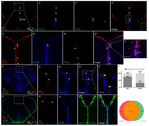
mdka is mainly expressed in non-proliferating RGCs in the telencephalon (A–A‴,B–B‴). Fluorescent in situ hybridization (FISH) with a probe directed against mdka mRNA (red) combined with Immunofluorescence (IF) with antibodies against PCNA (green) and S100β (blue) on cross-sections of WT telencephala. mRNA expression is detected in S100β+ cells and not detected in cells which are positive for PCNA. White arrows indicate mdka+/S100β+ cells. Yellow arrows indicate mdka−/PCNA+ cells. White arrowheads point to RMS. (A‴) inset: magnified view of the RMS, showing no co-expression between mdka mRNA and PCNA. Boxed-in area in (A) represents area of magnification in (B–B‴). Boxed-in area in (B‴) represents area of magnification in (C), showing two radial glial cells with their characteristic triangular shape and high expression of mdka. (D–E) FISH against mdka mRNA (red) with IF against the neuronal marker HuC/D (blue) indicating that mdka is not expressed in mature neurons since there is no co-localization between the two signals. Boxed-in area in (D) represents area of magnification in (D′–D‴). Boxed-in area in (D‴) represents area of magnification in (E). White arrowheads indicate mdka+/HuC/D− cells. (F) Quantification of S100β+, PCNA− type I and S100β+, PCNA+ type II cells expressing mdka mRNA. (G–H‴) FISH against mdka mRNA (red) combined with IF with antibodies against S100β (blue) on brains of the Tg(id1-CRM2:gfp) transgenic line (GFP, green). Expression of mdka mRNA is highly co-localized with cells positive for the transgene (green). White arrows show mdka+/id1-CRM2+ cells. Boxed-in area in (G) represents area of magnification in (H–H‴). (I) Quantification of cells expressing mdka mRNA in the Tg(id1-CRM2:gfp) transgenic line. Scale bar = 100 µm (A–A‴,D,G), 20 µm (B–C,D′–E,H–H‴). Location of cross-sections is indicated in lower right-hand corner of (A,D,G), respectively. n = 9 sections for quantifications. Abbreviations: id1, inhibitor of DNA binding 1; RMS, rostral migratory stream. EXPRESSION / LABELING:
|
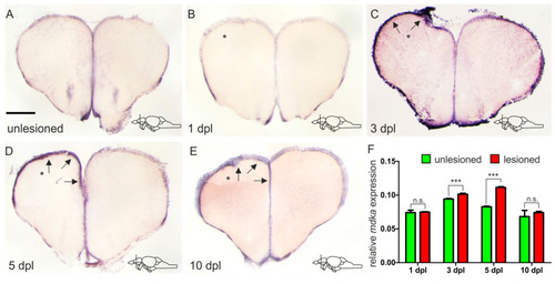
mdka is upregulated in a delayed fashion in response to injury. (A–E) ISH against mdka on cross-sections of the adult telencephalon in an unlesioned brain (A) and at different time points after the lesion (B–E). (B) Immediately after inflicting the lesion, there is no upregulation of mdka expression in the injured hemisphere. (C–E) mdka expression is increased in the injured hemisphere from 3 dpl onward (C) with a peak at 5 dpl (D), and then returns to baseline levels at 10 dpl (E). * The left, injured hemisphere is marked by an asterisk. Black arrows point at the expression of mdka in the ventricular zone of the left injured hemisphere. Location of cross-sections is indicated in the lower right-hand corner of (A–E). Scale bar = 100 µm. (F) RT-qPCR of mdka mRNA levels at different points after the lesion. Significance is indicated by asterisks: n.s. = not significant, *** p < 0.001. n = 3 brains/time point. Abbreviations: dpl, days post-lesion. |

After injury, mdka is still highly expressed in S100β+/PCNA− type I cells. (A–B‴) FISH against mdka mRNA (red) combined with IF using antibodies against S100β (blue) and PCNA (green) on telencephalic cross-sections at 5 dpl. After injury, mdka mRNA is highly co-expressed with S100β and not detected in cells which are positive for PCNA. Boxed-in area in (A) represents area of magnification in (B–B‴). White arrows point to mdka+/S100β+ cells. Yellow arrows show mdka−/PCNA+ cells. (C) Quantification of S100β+, PCNA− type I and S100β+, PCNA+ type II cells expressing mdka mRNA after injury. (D–E‴) FISH against mdka mRNA (red) combined with IF using antibodies against S100β (blue) and GFP (green) on brains of the Tg(id1-CRM2:gfp) transgenic line at 5 dpl. The signal for mdka mRNA is strongly co-localized with cells positive for the transgene. Boxed-in area in (D) represents area of magnification in (E–E‴). White arrows indicate mdka+/S100β+/id1+ cells. (F) Quantification of cells expressing mdka mRNA in the Tg(id1-CRM2:gfp) transgenic line at 5 dpl. The injured hemisphere is indicated with an asterisk. Scale bar = 100 µm (A,D), 20 µm (B–B‴,E–E‴). Location of cross-sections is indicated in (A,D), respectively. n = 9 sections for quantifications. |