|
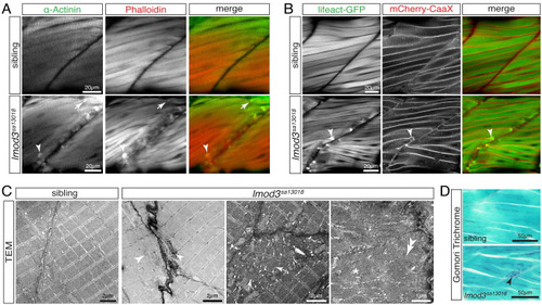
Aggregates within 4-dpf-old <italic toggle='yes'>lmod3</italic><sup><italic toggle='yes'>sa13018</italic></sup> are reminiscent of nemaline bodies formed within nemaline myopathy patients.(A) Immunohistochemistry using antibodies against α-Actinin (green) and phalloidin staining (red) marked aggregates located close to vertical myosepta of 4-dpf-old lmod3sa13018 homozygotes. Detected aggregates (arrowhead) were of various sizes and shapes, including rod-shaped structures (arrow). (B) Labelling of the myofibril with Lifeact-GFP (green) and the sarcolemma with mCherry-CaaX (red) confirmed aggregate (arrowhead) formation in lmod3sa13018 homozygotes at 4 dpf, again exclusively at vertical myosepta. (C) At 4 dpf, transmission electron micrographs (TEM) of skeletal muscle from siblings showed the typical myofibril striation and well-aligned sarcomeres. Comparable sarcomeres were also present within lmod3sa13018 homozygotes. However, close to the vertical myosepta, filament deposits reminiscent of fingerprint bodies (star), misaligned sarcomeres (arrows), and electron-dense aggregates of various sizes (arrowheads) were detected. (D) At 5 pdf, Gomori trichrome-stained sagittal sections depicted blue/purple structures close to the vertical myosepta within myofibres of lmod3sa13018 homozygotes. Scale bar sizes are indicated.
|