|
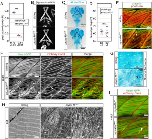
Thin filament deposits and myofibril disruptions lead to muscle weakness within the trunk but not head musculature of <italic toggle='yes'>capza1b</italic><sup><italic toggle='yes'>ex5</italic></sup> mutants.(A) Whereas 6-dpf-old siblings were able to generate a peak active force of 0.76 ± 0.01 mN, the force generated by capza1bex5 homozygotes was significantly reduced to 0.14 ± 0.01 mN (n = 4). (B) GFP fluorescence driven by Tg(-503unc:GFP) showed that the two hyohyoideus (hh) muscles of capza1bex5 homozygotes and siblings are comparable at 4 dpf (Z-stack projections of ventral views). (C) Focus stacks of Alcian Blue cartilage stains of capza1bex5 homozygotes and siblings were comparable at 7 dpf. (D) The ceratohyal angle (dotted lines) of capza1bex5 homozygotes was with 62 ± 2° not significantly different from the angle of 63 ± 1° formed in siblings (n = 6). (E) Phalloidin (red) and antibodies against α-Actinin (green) marked aggregates (arrowhead) close to the vertical myosepta within 3-dpf-old capza1bex5 homozygotes but not siblings. (F) Lifeact-GFP (green) highlighted organised myofibril (arrow) in life siblings and capza1bex5 homozygotes at 3dpf. In addition to the residual striation, capza1bex5 homozygotes featured myofibril ruptures (arrowhead) as well as aggregates and thin filament deposits at the peripheral myofibre ends. Transgenic mCherry-CaaX (red) was used to mark the sarcolemma and t-tubules. (G) On 3 dpf sagittal sections, Gomori trichrome-staining exposed blue/purple structures (arrowhead) close to the vertical myosepta of capza1bex5 homozygotes, but not siblings. (H) At 3 dpf, sarcomeres were organised within siblings as revealed by transmission electron micrographs. Organised sarcomeres were also present within capza1bex5 homozygotes (star). However, sarcomere organisation was lost in capza1bex5 close to myofibres’ ends and filament deposits, where isolated sarcomeric structures (arrow) and electron-dense aggregates (arrowhead), often with a lattice structure, were found instead. (I) Interestingly, myofibril ruptures were detected in 4-dpf-old capza1bex5/+ heterozygotes but not WT siblings, demonstrating capza1b haploinsufficiency. Data are presented as mean ± SEM; n.s. not significant and *** P < 0.001 calculated by Student’s t test. Scale bar sizes are indicated.
|