|
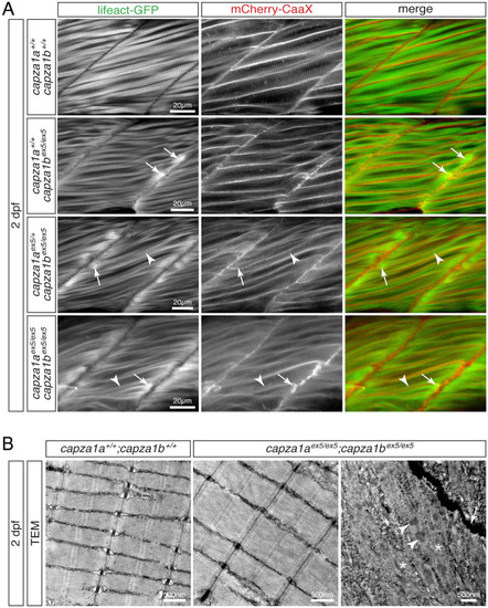
Residual sarcomeres assemble within <italic toggle='yes'>capza1a</italic> and <italic toggle='yes'>capza1b</italic> depleted zebrafish.(A) At 2 dpf, Lifeact-GFP (green) and mCherry-CaaX (red) highlighted the sarcomere organisation and myofibril striation within muscle fibres of WT siblings. In two out of four analysed capza1bex5 homozygotes, thin filament deposits (arrows) were detected at the peripheral ends of some fibres. Although myofibril striation (arrowheads) was detected in capza1bex5 homozygotes that were hetero- or homozygous for capza1aex5, thin filament deposits (arrows) were frequently located at the peripheral myofiber ends. (B) At 2 dpf, highly organised sarcomeres were found on transmission electron micrographs of siblings and capza1aex5;capza1bex5 compound homozygotes. However, electron-dense aggregates with a lattice structure (arrowheads) and filament deposits (star) were present close to the myosepta (arrow) in the compound homozygotes. Scale bar sizes are indicated.
|