IMAGE

Fig 3

ID
ZDB-IMAGE-220227-25
Genes
Antibodies
Source
Figures for Berger et al., 2022
Image
Figure Caption

Fig 3 Aggregates within 4-dpf-old <italic toggle='yes'>lmod3</italic><sup><italic toggle='yes'>sa13018</italic></sup> are reminiscent of nemaline bodies formed within nemaline myopathy patients.

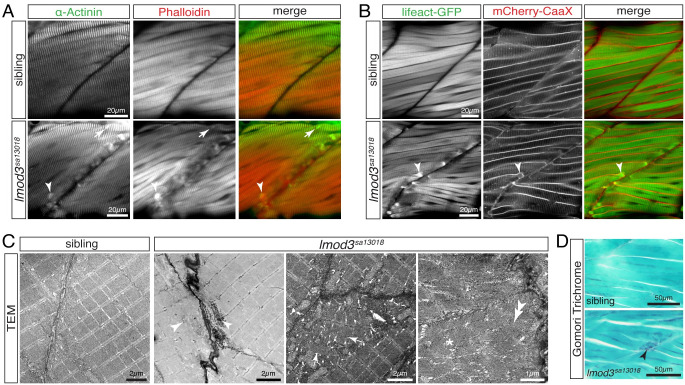
(A) Immunohistochemistry using antibodies against α-Actinin (green) and phalloidin staining (red) marked aggregates located close to vertical myosepta of 4-dpf-old lmod3sa13018 homozygotes. Detected aggregates (arrowhead) were of various sizes and shapes, including rod-shaped structures (arrow). (B) Labelling of the myofibril with Lifeact-GFP (green) and the sarcolemma with mCherry-CaaX (red) confirmed aggregate (arrowhead) formation in lmod3sa13018 homozygotes at 4 dpf, again exclusively at vertical myosepta. (C) At 4 dpf, transmission electron micrographs (TEM) of skeletal muscle from siblings showed the typical myofibril striation and well-aligned sarcomeres. Comparable sarcomeres were also present within lmod3sa13018 homozygotes. However, close to the vertical myosepta, filament deposits reminiscent of fingerprint bodies (star), misaligned sarcomeres (arrows), and electron-dense aggregates of various sizes (arrowheads) were detected. (D) At 5 pdf, Gomori trichrome-stained sagittal sections depicted blue/purple structures close to the vertical myosepta within myofibres of lmod3sa13018 homozygotes. Scale bar sizes are indicated.

Figure Data
Acknowledgments
This image is the copyrighted work of the attributed author or publisher, and ZFIN has permission only to display this image to its users. Additional permissions should be obtained from the applicable author or publisher of the image. Full text @ PLoS Genet.