
Fig. 6
- ID
- ZDB-FIG-220204-14
- Publication
- Hong et al., 2020 - In Situ Fucosylation of the Wnt Co-receptor LRP6 Increases Its Endocytosis and Reduces Wnt/β-Catenin Signaling
- Other Figures
- All Figure Page
- Back to All Figure Page
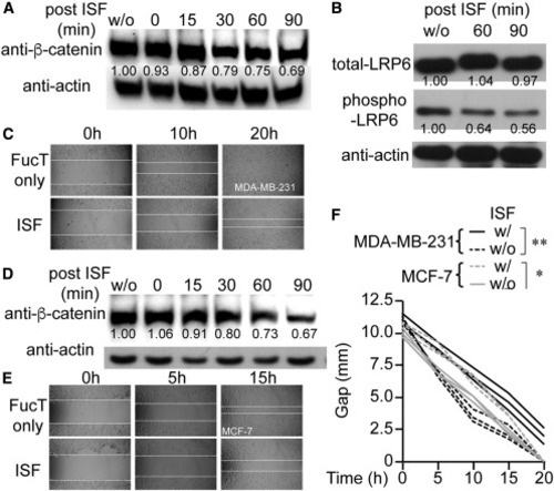
(A) Time-dependent decrease of the β-catenin level in ISF-treated MDA-MB-231. Western blot of anti-β-catenin (upper panel) and anti-actin (lower panel). (B) Immunoprecipitation of LRP6 from indicated MDA-MB-231 cells and detection of LRP6 and phosphorylated LRP6 by immunoblot. ISF-dependent decrease of LRP-6 phosphorylation. (C) Wound-healing assay results from MDA-MB-231 cells treated with ISF or not. (D) Time-dependent decrease of the β-catenin level in ISF-treated MCF-7. (E) Wound healing of MCF-7 cells treated with ISF or not. (F) Time-dependent gap closure of MDA-MB-231 and MCF-7 cells treated with ISF or not in wound-healing assays. One-way ANOVA: ∗p < 0.05, ∗∗p < 0.01. In (A), (B), and (D), the number indicates the relative protein level normalized to the corresponding level of actin. |