
Fig. 4
- ID
- ZDB-FIG-220204-12
- Publication
- Hong et al., 2020 - In Situ Fucosylation of the Wnt Co-receptor LRP6 Increases Its Endocytosis and Reduces Wnt/β-Catenin Signaling
- Other Figures
- All Figure Page
- Back to All Figure Page
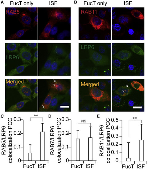
(A and B) Confocal visualization of LRP6 (FLAG-LRP6, green) in FucT only-treated and ISF-treated Pro-5 cells. Early endosome marker RAB5-mCherry (A, red) and recycling endosome marker RAB11-mCherry (B, red) were transiently expressed in Pro-5 cells. The co-localization of LRP6 with RAB5 or RAB11 is shown in yellow. The white arrows indicate the presentative co-localization of LRP6 with RAB5 or RAB11, respectively. Scale bars, 20 μm. (C–E) Co-localization of RAB5 (C), RAB7 (D), or RAB11 (E) with LRP6 in FucT-only-treated and ISF-treated Pro-5 cells, is shown as Pearson's correlation coefficient. Two-sided Student's t test: ∗∗p < 0.01; NS, not significant. |