
Figure 5
- ID
- ZDB-FIG-220104-70
- Publication
- Tobia et al., 2021 - An Orthotopic Model of Uveal Melanoma in Zebrafish Embryo: A Novel Platform for Drug Evaluation
- Other Figures
- All Figure Page
- Back to All Figure Page
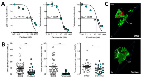
Effect of anticancer drugs on the growth of human uveal melanoma 92.1-RFP+/luc+ xenografts. (A) Effect of paclitaxel, panobinostat, and everolimus treatments on the proliferation of 92.1-RFP+/luc+ cells in vitro. Viable cells were counted after 48 h of incubation with increasing concentrations of paclitaxel or panobinostat or after 72 h of incubation with everolimus. Data are the mean ± SEM of two independent experiments. (B) After 92.1-RFP+/luc+ cell grafting into the zebrafish eye, 0.4 pmoles/embryo of paclitaxel, panobinostat, everolimus or the corresponding volume of DMSO were injected in the same eye. Tumor growth was evaluated at t4 by measuring the cell luminescence signal in the lysates of the whole embryos. Data are the mean ± SEM of two independent experiments. Each dot represents one embryo. * p < 0.05 and *** p < 0.001 vs. DMSO, Student’s t-test. (C) 3D reconstruction of the eye region of 92.1-RFP+/luc+ xenografts evaluated 4 days post implantation in the absence or in the presence of paclitaxel injection. Scale bar: 50 µm. Asterisk indicates the superficial ocular vasculature; CVP, choroidal vascular plexus. |