
Figure 1
- ID
- ZDB-FIG-220104-66
- Publication
- Tobia et al., 2021 - An Orthotopic Model of Uveal Melanoma in Zebrafish Embryo: A Novel Platform for Drug Evaluation
- Other Figures
- All Figure Page
- Back to All Figure Page
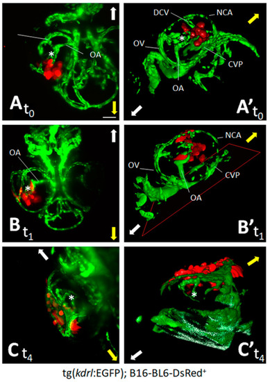
Zebrafish embryo eye is a microenvironment suitable for cell grafting. Murine melanoma B16-BL6-DsRed+ cells (100 cells/embryo) were orthotopically injected in the posterior side of the developing eye of transgenic tg(kdrl:EGFP) zebrafish embryos at 48 hpf. Maximum intensity projection of the z-stacks (A–C) and 3D reconstructions (A’–C’) of B16-BL6-DsRed+ cells performed at 1 h (t0) (A,A’), 1 day (t1) (B,B’), and 4 days (t4) (C,C’) post implantation. (A,B) ventral view; (C) dorsal view. Asterisk indicates the hyaloid artery. Arrows indicate embryo orientation: white arrow, posterior side; yellow arrow, anterior side. CVP, choroidal vascular plexus; DCV, dorsal ciliary vein; NCA, nasal ciliary artery; OA, optic artery; OV, optic vein. Scale bar: 50 µm. |