
Figure 3
- ID
- ZDB-FIG-220104-68
- Publication
- Tobia et al., 2021 - An Orthotopic Model of Uveal Melanoma in Zebrafish Embryo: A Novel Platform for Drug Evaluation
- Other Figures
- All Figure Page
- Back to All Figure Page
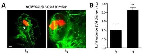
Luciferase-based quantification of the growth of human melanoma A375M-RFP+/luc+ xenografts. Human melanoma A375M-RFP+/luc+ cells (100 cells/embryo) were injected into the posterior side of the developing eye of transgenic tg(kdrl:EGFP) zebrafish embryos at 48 hpf. (A) Maximum intensity projection of the z-stacks of A375M-RFP+/luc+ cells performed at 1 h (t0) and 4 days (t4) post implantation. T0, lateral view, anterior to the top; t4, dorsal view, anterior to the top. Asterisk indicates the superficial ocular vasculature. Scale bar: 50 µm. (B) Evaluation of A375M-RFP+/luc+ bioluminescence signal in the lysates of the whole embryos at t0 and t4. Data are the mean ± SEM (n = 8). ** p < 0.01 vs. t0, Student’s t-test. |