
Figure 5—figure supplement 2.
- ID
- ZDB-FIG-211029-175
- Publication
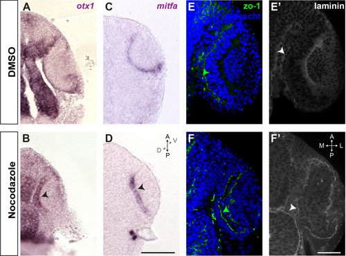
- Moreno-Mármol et al., 2021 - Stretching of the retinal pigment epithelium contributes to zebrafish optic cup morphogenesis
- Other Figures
-
- Figure 1—video 1.
- Figure 2—source data 1.
- Figure 3—figure supplement 1—source data 1.
- Figure 3—figure supplement 1—source data 1.
- Figure 4—source data 1.
- Figure 5—figure supplement 1.
- Figure 5—figure supplement 1.
- Figure 5—figure supplement 2.
- Figure 6—source data 1.
- Figure 7—figure supplement 1.
- Figure 7—figure supplement 1.
- Figure 8.
- All Figure Page
- Back to All Figure Page
( |