FIGURE

FIGURE 2

ID
ZDB-FIG-211009-9
Publication
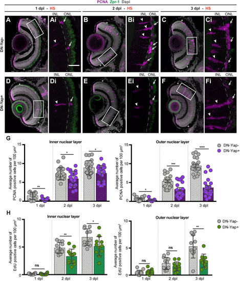
Lourenço et al., 2021 - Yap Regulates Müller Glia Reprogramming in Damaged Zebrafish Retinas
Other Figures
All Figure Page
Back to All Figure Page
FIGURE 2

Yap inhibition reduces cell proliferation in the retina after photoreceptor-induced light lesion. (A–F) Transverse cryosections of DN-Yap− (A–C) and DN-Yap+ (D–F) retinas immunostained for proliferating cell nuclear antigen (PCNA) (magenta) and Zpr-1 (green) and counterstained with DAPI (gray). White arrowheads and arrows indicate PCNA+ cells in the inner nuclear layer (INL) and outer nuclear layer (ONL), respectively, at 1 dpl (A,D magnified, Ai,Di), 2 dpl (B,E, magnified, Bi,Ei), and 3 dpl (C,F, magnified, Ci,Fi). (G) Quantification of PCNA + cells in DN-Yap− and DN-Yap + larva retinas from 1 to 3 dpl, in the INL and ONL. (H) Quantification of EdU + cells in DN-Yap− and DN-Yap + larva retinas from 1 to 3 dpl, in the INL and ONL. ns, non-significant; *p < 0.05, **p < 0.01, ***p < 0.001, ****p < 0.0001. Unpaired t-test with Welch’s correction. Asterisks delimitate the lesioned region. Dashed lines delimitate INL from ONL. White boxes delimitate magnified (Ai–Fi). Scale bars correspond to 50 μm in (A,C), and 20 μm in magnified (Ai,Ci).

Expression Data

Expression Detail
Antibody Labeling
Phenotype Data

Phenotype Detail
Acknowledgments
This image is the copyrighted work of the attributed author or publisher, and ZFIN has permission only to display this image to its users. Additional permissions should be obtained from the applicable author or publisher of the image. Full text @ Front Cell Dev Biol