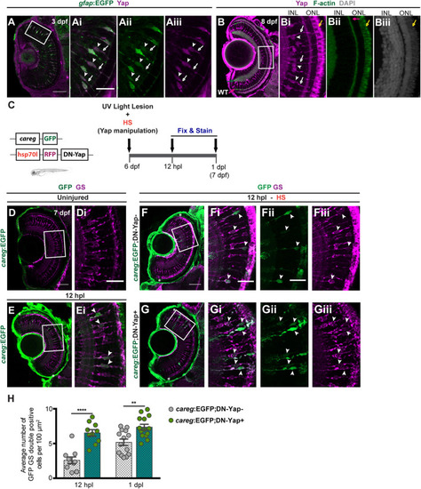
Yap inhibition induces accumulation of activated Müller glial cells (MGs) upon photoreceptor-induced light lesion. (A) Transverse cryosection of a Tg(gfap:GFP) 3-dpf retina immunostained for green fluorescent protein (GFP) (green, Ai) and Yap (magenta, Aii), indicating Yap localization in MG cell bodies (arrowheads) and radial processes (arrows) (Ai–Aiii). (B) Transverse cryosection of a WT 8-dpf retina immunostained for Yap (magenta, B,Bi) and F-actin (green, B,Bii) and counterstained with DAPI (gray, Biii), indicating photoreceptors outer-segment autofluorescence (yellow arrow). (C) Schematic representation of the UV light lesion assay. (D,E) Transverse cryosections of uninjured 7-dpf careg-EGFP (D,Di) and 12-hpl careg-EGFP (E,Ei) larva retinas immunostained for GFP (green) and glutamine synthetase (GS) (magenta). White arrowheads indicate GFP-positive MGs activating careg (E, magnified, Ei). (F,G) Transverse cryosections of 12-hpl careg:EGFP;DN-Yap− controls (F-Fiii) and careg:EGFP;DN-Yap+ (G-Giii) retinas immunostained for GFP (green) and GS (magenta). White arrowheads indicate GFP-positive MGs activating careg. (H) Quantification of the number of double GFP GS-positive cells in careg:EGFP;DN-Yap− controls and careg:EGFP;DN-Yap + larva retinas from 12 hpl to 1 dpl. **p < 0.01, ****p < 0.0001; unpaired t-test with Welch’s correction. White boxes delimitate magnified (Ai–Giii). Green arrow indicates the inner plexiform layer. Pink arrow indicates the outer plexiform layer. Scale bars correspond to 50 μm in (A,D,F) and 20 μm in magnified (Ai,Di,Fi). Asterisks delimitate the lesioned region.
|
