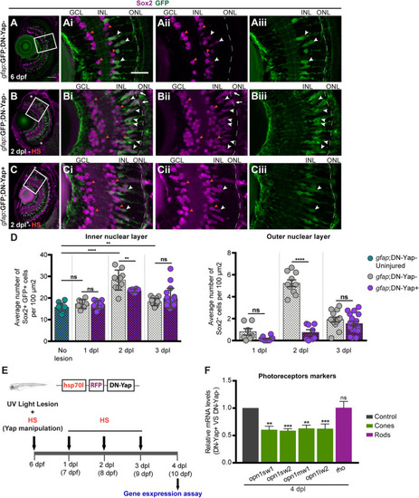
Yap is required to regulate the number of Sox2-positive Müller glia cell (MG) progenitor cells and expression of photoreceptor markers after photoreceptor-induced light lesion. (A–C) Transverse cryosections of uninjured 6-dpf gfap:GFP;DN-Yap− control retina (A), 2-dpl gfap:GFP;DN-Yap− (B) and gfap:GFP;DN-Yap+ (C) retinas subjected to UV light lesion and heat shock (HS) at 6 dpf, immunostained for Sox2 (magenta) and green fluorescent protein (GFP) (green). Sox2 is localized in amacrine cells (ACs) (orange arrowheads) (magnified, Ai,Aii,Bi,Bii,Ci,Cii), MGs (white arrowheads), and progenitors in the outer nuclear layer (ONL) (white arrows) (magnified, Ai–Aiii,Bi–Biii,Ci–Ciii). (D) Quantification of Sox2 + GFP + cells in gfap:GFP;DN-Yap− uninjured controls at 6 dpf, and lesioned gfap:GFP;DN-Yap− and gfap:GFP;DN-Yap + larva retinas from 1 to 3 dpl, in inner nuclear layer (INL) and ONL. (E) Schematic representation of the UV light lesion and HS assay. (F) Relative gene expression of photoreceptor markers at 4 dpl (n = 7 biological replicates) in DN-Yap + versus DN-Yap− controls. ns, non-significant; **p < 0.01, ***p < 0.001, ****p < 0.0001. Unpaired t-test with Welch’s correction for Sox2+ cell quantification. Paired t-test for the relative gene expression of photoreceptor markers. Asterisks delimitate the lesioned region. Dashed lines delimitate INL from ONL. White boxes delimitate magnified (Ai–Aiii,Bi–Biii,Ci–Ciii). Scale bars correspond to 50 μm in (A) and 20 μm in magnified (Ai).
|
