
FIGURE
FIGURE 6
- ID
- ZDB-FIG-210611-54
- Publication
- Morris et al., 2021 - A Novel Lysolecithin Model for Visualizing Damage in vivo in the Larval Zebrafish Spinal Cord
- Other Figures
- All Figure Page
- Back to All Figure Page
FIGURE 6
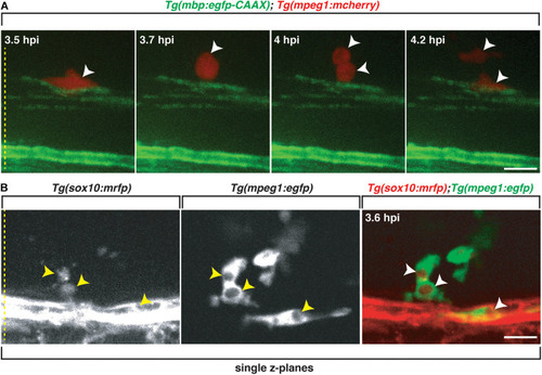
Professional phagocytes proliferate and clear Sox10+ debris. All images are lateral views of the spinal cord with anterior to the left and dorsal to the top. |
Expression Data
| Genes: | |
|---|---|
| Fish: | |
| Condition: | |
| Anatomical Terms: | |
| Stage: | Day 6 |
Expression Detail
Antibody Labeling
Phenotype Data
| Fish: | |
|---|---|
| Condition: | |
| Observed In: | |
| Stage: | Day 6 |
Phenotype Detail
Acknowledgments
This image is the copyrighted work of the attributed author or publisher, and
ZFIN has permission only to display this image to its users.
Additional permissions should be obtained from the applicable author or publisher of the image.
Full text @ Front Cell Dev Biol