
Image
|
Figure Caption
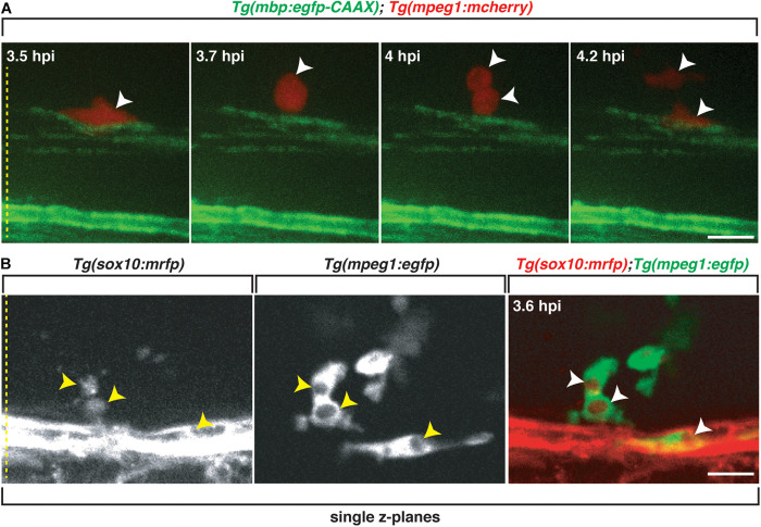
FIGURE 6
Professional phagocytes proliferate and clear Sox10+ debris. All images are lateral views of the spinal cord with anterior to the left and dorsal to the top.
Figure Data
Acknowledgments
This image is the copyrighted work of the attributed author or publisher, and
ZFIN has permission only to display this image to its users.
Additional permissions should be obtained from the applicable author or publisher of the image.
Full text @ Front Cell Dev Biol