
Fig. 4
- ID
- ZDB-FIG-210510-20
- Publication
- Barker et al., 2020 - Functional, molecular and morphological heterogeneity of superficial interneurons in the larval zebrafish tectum
- Other Figures
- All Figure Page
- Back to All Figure Page
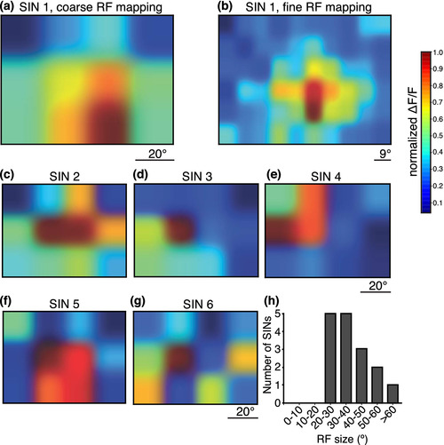
Gal4s1156t+ SINs have large receptive fields. Receptive fields of SINs were mapped using a 12‐square grid stimulus. White squares corresponding to 20° of the larva's visual field were flashed over the whole visual field on a black background. (a) An example SIN RF. (b) Finer mapping using a 60‐square grid for the same cell in (a) revealed a similar RF map. (c–g). Five additional examples of SIN RFs mapped with the 12‐square grid stimulus. Most cells had a central area of maximal activity (c–f). (h) RF areas for 26 Gal4s1156t+ SINs are plotted here. All RFs were greater than 20° of the larva's visualfield |