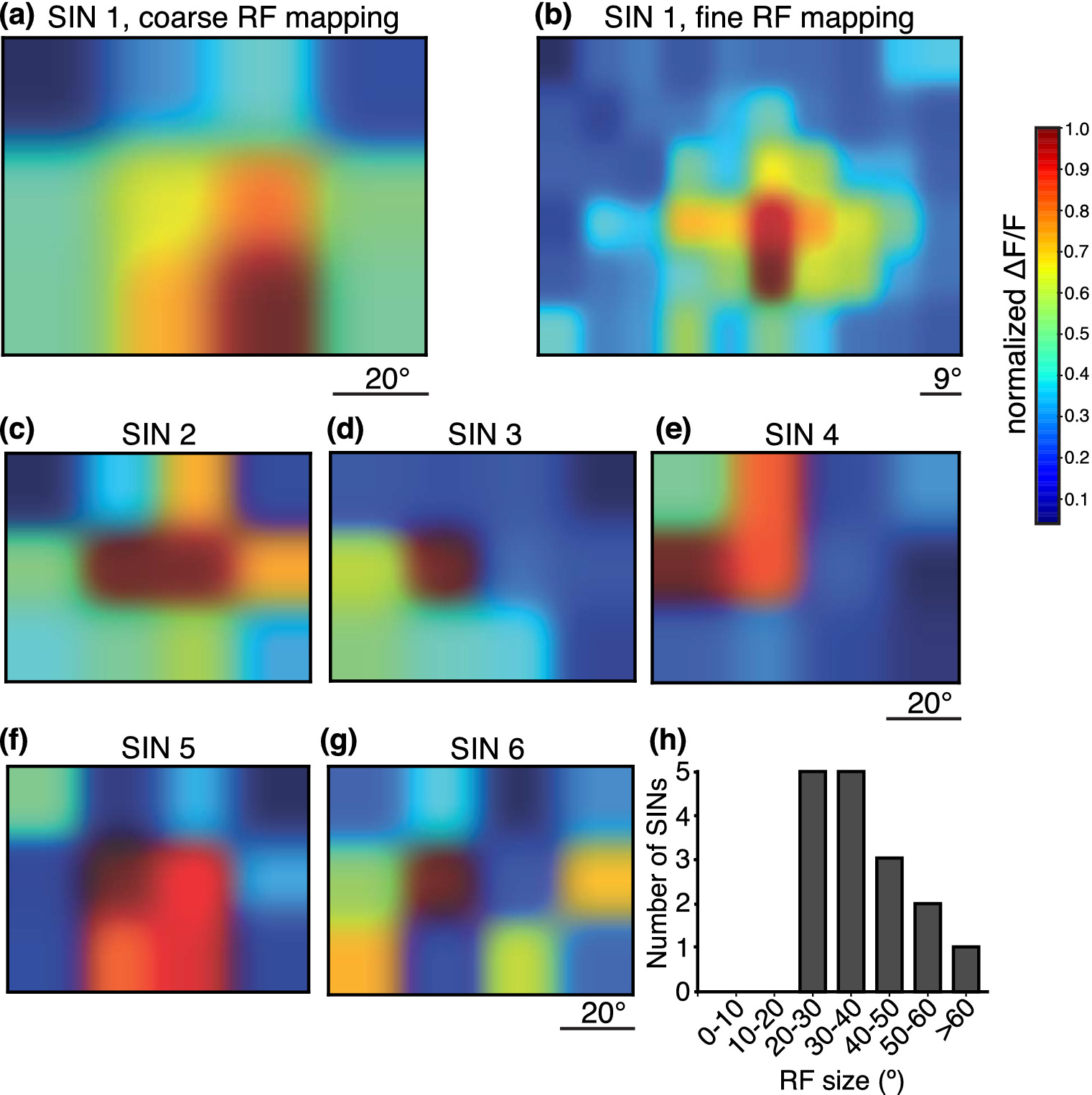
Fig. 4 Gal4s1156t+ SINs have large receptive fields. Receptive fields of SINs were mapped using a 12‐square grid stimulus. White squares corresponding to 20° of the larva's visual field were flashed over the whole visual field on a black background. (a) An example SIN RF. (b) Finer mapping using a 60‐square grid for the same cell in (a) revealed a similar RF map. (c–g). Five additional examples of SIN RFs mapped with the 12‐square grid stimulus. Most cells had a central area of maximal activity (c–f). (h) RF areas for 26 Gal4s1156t+ SINs are plotted here. All RFs were greater than 20° of the larva's visualfield
Image
Figure Caption
Acknowledgments
This image is the copyrighted work of the attributed author or publisher, and
ZFIN has permission only to display this image to its users.
Additional permissions should be obtained from the applicable author or publisher of the image.
Full text @ J. Comp. Neurol.

